Gefragt von: Jared Korinko
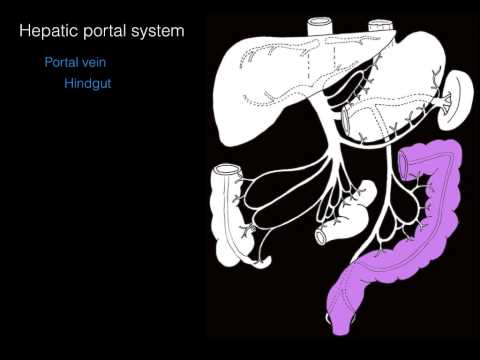
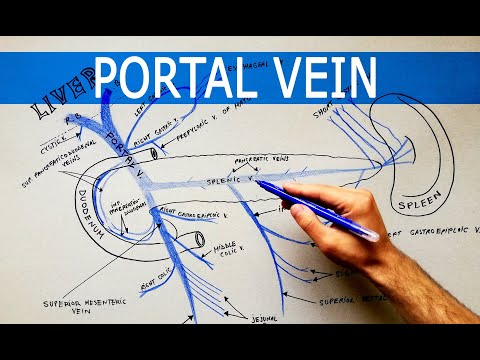
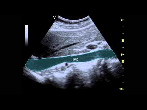
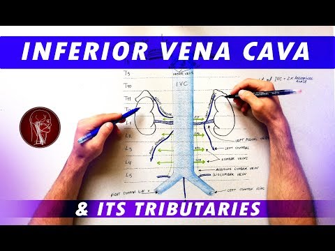
Fragesteller Allgemeines

Ivc Portal Vein Anatomy

Der Link der Ivc Portal Vein Anatomy-Seite ist unten angegeben. Seiten, die sich auf Ivc Portal Vein Anatomy beziehen, werden ebenfalls aufgelistet.

Zuletzt aktualisiert: 2021-04-19

11

Added by: Christopher Kroek

Erklärer

Portal vein | Radiology Reference Article | …

Gross anatomy. The portal vein usually measures approximately 8 cm in length in adults with a maximum diameter of 13 mm. It originates posterior to the neck of the pancreas where it is classically formed by the union of the superior mesenteric and splenic veins (the portovenous confluence). The origin of the vein defines the location of the pancreatic neck.
Url: https://radiopaedia.org/articles/portal-vein
Portal vein | Radiology Reference Article | …

8,039,278

Monatliche Besuche

12,108

Alexa Rank

GB

Beliebt in

Up

Service Status

Added by: Christine Flanagan

Erklärer

Enlarged azygos vein | Radiology Reference …

Terminology. Spelling it "azygous" when referring to the vein is incorrect, regardless of whether British or American English 5.Azygous is a word in English meaning 'without pair', but in the context of anatomy, see Terminologia Anatomica, azygos vein is the sole correct spelling.
Url: https://radiopaedia.org/articles/enlarged-azygos-vein
Enlarged azygos vein | Radiology Reference …

8,039,278

Monatliche Besuche

12,108

Alexa Rank

GB

Beliebt in

Up

Service Status

Added by: Narelle Mills

Erklärer

Inferior Vena Cava - Anatomy Pictures and …

15.12.2016 · The hepatic vein provides blood from the digestive organs of the abdomen after it has passed through the hepatic portal system in the liver. Blood from the tissues of the lower back, including the spinal cord and muscles of the back, enters the vena cava through the lumbar veins. Many smaller veins also provide blood to the vena cava from the tissues of the abdominal body wall. Upon reaching ...
Url: https://www.innerbody.com/image_dige07/card26.html
Inferior Vena Cava - Anatomy Pictures and …

1,283,531

Monatliche Besuche

74,741

Alexa Rank

US

Beliebt in

Up

Service Status

Added by: Andrew Crosbie

Erklärer

Portal Hypertension: Practice Essentials, …

30.11.2017 · Portal vein and associated anatomy. View Media Gallery. Previous Next: ... Presinusoidal venous block (eg, portal vein thrombosis, schistosomiasis, primary biliary cirrhosis) - Characterized by elevated portal venous pressure and a normal wedged hepatic venous pressure (WHVP); these abnormalities cannot be detected by surrogate measurement (WHPV, HVPG), because the measured …
Url: https://emedicine.medscape.com/article/182098-overview
Portal Hypertension: Practice Essentials, …

29,242,425

Monatliche Besuche

3,363

Alexa Rank

US

Beliebt in

Up

Service Status

Added by: Jako Ebersohn

Erklärer

Inferior vena cava: Anatomy and function | …

08.04.2021 · The inferior vena cava (IVC) is the largest vein of the human body. It is located at the posterior abdominal wall on the right side of the aorta. The IVC’s function is to carry the venous blood from the lower limbs and abdominopelvic region to the heart.. The inferior vena cava anatomy is essential due to the vein’s great drainage area, which also makes it a hot topic for anatomy exams.
Url: https://www.kenhub.com/en/library/anatomy/inferior-vena-cava
Inferior vena cava: Anatomy and function | …

3,021,920

Monatliche Besuche

31,962

Alexa Rank

US

Beliebt in

Up

Service Status

Added by: Arthur Sinclair

Erklärer

The Radiology Assistant : Segmental Anatomy

The portal vein divides the liver into upper and lower segments. The left and right portal veins branch superiorly and inferiorly to project into the center of each segment. On a frontal view of the liver the posteriorly located segments VI and VII are not visible. The illustration above is a schematic presentation of the liver segments. In reality however the proportions are different. On a ...
Url: https://radiologyassistant.nl/abdomen/liver/segmental-anatomy
The Radiology Assistant : Segmental Anatomy

1,066,650

Monatliche Besuche

89,806

Alexa Rank

GB

Beliebt in

Up

Service Status

Added by: Michael Barrett

Erklärer

Duodenal Anatomy: Overview, Gross Anatomy, …

08.12.2017 · The first (superior) part, or duodenal bulb or cap (5 cm), which is connected to the undersurface of the liver (porta hepatis) by the hepatoduodenal ligament (HDL), containing the proper hepatic artery, portal vein, and common bile duct (CBD); the quadrate lobe (segment IV) of the liver and the gallbladder are in front, whereas the CBD, the portal vein (PV), and the gastroduodenal artery …
Url: https://emedicine.medscape.com/article/1898874-overview
Duodenal Anatomy: Overview, Gross Anatomy, …

29,242,425

Monatliche Besuche

3,363

Alexa Rank

US

Beliebt in

Up

Service Status

Added by: Kenhub - Learn Human Anatomy

Erklärer

Hepatic Portal Vein (preview) - Human Anatomy | Kenhub

Added by: The Noted Anatomist

Erklärer

Hepatic portal system

Added by: Sam Webster

Erklärer

Hepatic portal vein (anatomy)

Added by: Anatomy Knowledge

Erklärer

The PORTAL VEIN and its tributaries | Anatomy Tutorial

Added by: AnatomyZone

Erklärer

Blood Supply to the Gut (Introduction) - Part 2 (Venous Drainage/Portal System)

Added by: Taim Talks Med

Erklärer

Portal Venous System & Porto-Caval Anastomosis (Tributaries and Clinical Significance) - Anatomy

Added by: Smart Med Learn Dr. Ankit Jain

Erklärer

Portal vein / Anatomy / Simplified - Formation, Relations and Tributaries

Added by: Taim Talks Med

Erklärer

Inferior Vena Cava (Topography, Visceral and Parietal Tributaries) - Anatomy

Added by: Ultrasound at U of SC School of Medicine

Erklärer

Ultrasound of the IVC

Added by: Anatomy Knowledge

Erklärer

Inferior Vena Cava and its tributaries - Anatomy Tutorial